

Necrotising fasciitis after hysterectomy and concomitant transvaginal mesh repair in a patient with pelvic organ prolapse
Article authors


Clinic of Urology MSMSU
Abstract
Necrotising fasciitis is a severe form of soft tissue infection. Herein, we present an unreported complication of the transvaginal repair of a pelvic organ prolapse (POP) with trocar-guided polypropylene mesh and a concomitant hysterectomy. A 61-year-old Caucasian female who had been using an intrauterine device (IUD) for 30 years presented with a stage 3 pelvic organ prolapse. A genital ultrasound examination confirmed the presence of an IUD, but found no endometrial abnormalities. The surgical management was limited to a transvaginal hysterectomy and simultaneous anterior vaginal wall repair augmented with trocarguided mesh. A morphological examination of the removed uterus confirmed the presence of the intrauterine device and additionally found endometrial cancer (T1N0M0), which was not revealed during the preoperative ultrasound. Within 6 days of the surgery, she developed anaerobic bilateral necrotising fasciitis on both thighs. Non-clostridial streptococci were identified in the wound. After 18 days of intensive care, the patient died of fatal coagulopathy.
Keywords Necrotising fasciitis . Anaerobic . Intrauterine device . Prolapse . Mesh . Endometrial cancer
Introduction
Necrotising fasciitis is a sever e form of soft tissue infection. Herein, we present an unreported case of necrotising fasciitis of both thighs in a patient with pelvic organ prolapse, endometrial cancer and an intrauterine device. This patient was treated with hysterectomy and pelvic floor repair with synthetic mesh.
Case report
A 61-year-oldCaucasian woman presented withstage 3 pelvic organ prolapse, which was predominantly caused by cystocele, and urinary incontinence. According to her history, the patient used an intrauterine device (IUD) that was placed approximately 30 years ago for contraceptive purposes. A preoperative physical examination showed an anterior wall stage 3 (POP-Q) prolapse with significant uterine descent behind the hymenal ring. A vaginal examination showed no signs of the IUD removal string and no abnormal discharge from the cervix. A stress cough test was positive. Uterine ultrasound demonstrated the presence of the IUD and showed nohyperplasia ofthe endometrial tissue. She was not suffering with diabetes mellitus and her status was not immunocompromised. The patient opted for vaginal hysterectomy and prolapse repair with a mesh kit (Prolift Anterior, Gynecare; Ethicon). Anti-incontinence surgery was delayed as a second-step procedure, in accordance with in-hospital protocol. The surgery took 110 min to perform, and more than 500 cc of blood loss occurred intraoperatively. The patient was intravenously administered 2nd generation cephalosporin and metronidazole beginning on the day of surgery. A morphological examination after the surgery demonstrated the presence of the IUD and endometrial cancer (T1N0M0).
In the early postoperative period, the urethral catheter was removed, and proper voiding and bowel function were restored. The patient remained subfebrile with a normal blood cell count. On the 6th day after surgery, the patient developed swelling and redness on both thighs with severe pain, high fever, hypotension and other signs of systemic toxicity. A physical examination showed pigmentation on the medial parts of both thighs with gas crepitation (Fig. 1 ).
Necrotising fasciitis of both thighs was diagnosed. The patient was immediately taken to the surgical theatre for fasciotomy and debridement of the affected areas (Fig. 2 ). There were no signs of retroperitoneal pelvic inflammation or involvement of the obturator areas or the synthetic prosthesis in the anaerobic process. Intravenous imipenem, 500 mg qid, was started immediately. Non-clostridial streptococci were identified in the wound. After surgery, the patient was treated in the intensive care unit with two additional surgical debridements over the next 18 days. Unfortunately, the patient died as a result of fatal coagulopathy.
Discussion
Necrotising fasciitis is a severe form of soft tissue infection [ 1 ]. It is characterised by widespread fascial necrosis that is primarily caused by Streptococcus hemolyticus or Clostridium perfringens . After the onset of the disease, patients typically become colonised w ith their own aerobic and anaerobic microflora from the gastrointestinal or urogenital tracts. In women, Bartholin abscesses and vulval skin infections may cause necrotising fasciitis. The infection is typically associated with immunocompromised patients, but several cases have also been associated with endometrial cancer. At the present time, this issue is limited to only several case reports [ 2 ].
Before abortion was legalised in most countries, anaerobic infections were commonly reported in gynaecological patients and often had devastating consequences. This may be due to the haematogenous spread of an infection from the uterus. The presence of an intrauterine device may enhance the risk of endogenous infection. In IUD users, the combinations of several anaerobic/aerobic microbes were associated with an increased risk of inflammatory complications, irrespective of the duration of IUD use. Long-term IUD use appeared to be associated with an increased risk of a pelvic inflammatory disease being complicated [ 3 ]
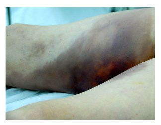
Fig. 1 Pigmentation on the medial parts of the thighs with gas crepitation
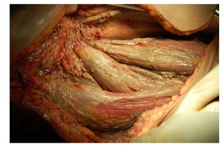
Fig. 2 Fasciotomy and debridement of the affected areas were performed
Recently, we have witnessed the widespread use of synthetic materials in female pelvic floor reconstruction and in the treatment of stress urinary incontinence. Several case reports have described necrotising fasciitis after the insertion of synthetic tape [ 4 ]. Most likely, the infection was due to injuries to the integumentary system in the area of mesh insertion. In our particular case, we found no direct involvement of the mesh or trocar passage areas in the anaerobic process. Surgical management includes a wide incision and the debridement of all involved areas followed by intensive care and antibiotics. Even with modern approaches, the mortality of necrotising fasciitis remains high and ranges from 11 to 45 %, despite improvements in critical care, the use of broad-spectrum antibiotics and aggressive surgical debridement [ 5 ].
Conclusion
To our knowledge, this is the first report to describe a case of necrotising fasciitis of the lower limbs in a patient with pelvic organ prolapse, endometrial cancer and an intrauterine device, who was treated with hysterectomy and pelvic floor repair with synthetic mesh. This case should warn pelvic floor surgeons to consider patients with IUDs and endometrial cancer as high-risk patients (grade IV level of evidence), and the use of synthetic materials in these patients should be limited.
References
1. Hung CC, Chang SC, Lin SF et al (1996) Clinical manifestations, microbiology and prognosis of 42 patients with necrotizing fasciitis. J Formos Med Assoc 95:917 – 922
2. Braverman J, Adachi A, Lev-Gur M et al (1987) Spontaneous clostridia gas gangrene of uterus associated with endometrial malignancy. Am J Obstet Gynecol 156(5):1205 – 1207
3. Viberga I, Odlind V, Lazdane G, Kroica J, Berglund L, Olofsson S (2005) Microbiology profile in women with pelvic inflammatory disease in relation to IUD use. Infect Dis Obstet Gynecol 13 (4):183 – 190
4. Lee KY, Sim JA, Lee SW, Kim TB, Yoon SJ, Park KS, Kim KH (2011) Necrotizing fasciitis following transobturator tape procedure: a case report and literature review. Can Urol Assoc J 5(4):E65 – E68
5. Roje Z, Roje Z, Mati ć D, Librenjak D, Dokuzovi ć S, Varvodi ć J (2011) Necrotizing fasciitis: literature review of contemporary strategies for diagnosing and management with three case reports: torso, abdominal wall, upper and lower limbs. World J Emerg Surg 6 (1):46