

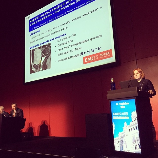
Novel method for evaluation of pelvic floor mobility in women with genital prolapse and stress urinary incontinence
Article authors


1 Moscow State University of Medicine and Dentistry, Dept. of Urology, Moscow, Russia
Introduction & Objectives
This study presents a new potentially useful three-dimensional non-invasive tool to study pelvic floor mobility in women.
Material & Methods
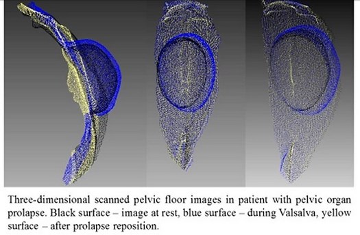
Patients suffering from pelvic organ prolapse (POP), stress urinary incontinence (SUI) and healthy volunteers (controls) were included in the study. The study was approved by the local Ethics committee. All participants were scanned at rest and during Valsalva maneuver using an Artec™ 3D portable scanner. Patients suffering from major-grade prolapse were also scanned after the repositioning of the POP. After the generation of the pelvic floor 3D model, the volume of the prolapsed vaginal wall was measured as dynamic prolapse increment (DPI), which was defined as a growth in the prolapse volume from rest to maximal Valsalva probe (DPI = (Vval – Vrest) / Vrest %).
Results
Overall, 72 women participated in this study. The mobility of the pelvic floor of women suffering from SUI was 1.6 times higher when compared with control group. Comparison of the subgroups of patients with POP revealed that the figure of 75% DPI was a threshold for the onset of symptoms POP, and abnormal mobility of the pelvic floor was marked with a 52% growth in prolapse volume when compared with the norm. For some patients with severe POP the increase in the prolapse volume was not related to the further descent of the most prominent point (Figure 1). Instead, it was caused by the enlargement of the other compartments, such as the cystocele or enterocele. That enlargement occurred in 3D manner and could not be validated by the POP-Q system.
Conclusions
The three-dimensional pelvic floor modeling reveals pathological mobility of the pelvic floor in the early stages of pelvic organ prolapse before its clinical manifestations that provides an opportunity for preventive measures and objectifies dynamic monitoring data. This technique also helps to determine the functional reserves of the pelvic floor mobility during planning surgery for pelvic dysfunction, particularly to prevent functional complications because of the prolapsed organs over fixation by mesh implantation.
The study was funded by the governmental grant, project МК-1921.2013.7